

Consulting
Consultation on the scientific research question
Decision-making on CLSM, EM, or a combined approach
Experimental design, including the selection of antibody panels as well as fixation and sample preparation strategies
Electron microscopy enables high-resolution analysis of cellular ultrastructure, providing detailed information on cell architecture, content, morphology, and elemental composition that is inaccessible to other imaging techniques. With resolution reaching the sub-nanometer scale, it remains essential for the investigation of subcellular compartments and organelles in basic and translational research. Through analytical electron microscopy, morphological information can be complemented by chemical characterization, including elemental analysis of nanoparticles. Integrated with fluorescence microscopy as a complementary approach, our core facility supports correlative workflows that bridge functional light microscopy and ultrastructural analysis across a wide range of medical and biological research applications.
Watch the video on Youtube
Consultation on the scientific research question
Decision-making on CLSM, EM, or a combined approach
Experimental design, including the selection of antibody panels as well as fixation and sample preparation strategies

Embedding of tissues or cells in Epon or Lowicryl
Semi-& ultrathin sectioning of embedded samples
Morphological analysis of embedded samples
EFTEM-based elemental analysis
Immunogold labelling of embedded samples
(Immuno-) Negativestaining

Preparation of Cryo- or Paraffinsections
Immunofluorescence staining
Confocal Laserscanning Imaging

Image and data analysis
Preparation of images to be ready for publication
Illustration of graphs and figures

Dr. Jule Riebelmann
Head of Core Facility Electron Microscopy
Prof. Dr. Martin Schaller
Deputy Head of Core Facility Electron Microscopy
Anneli Yilmaz
Technical Head
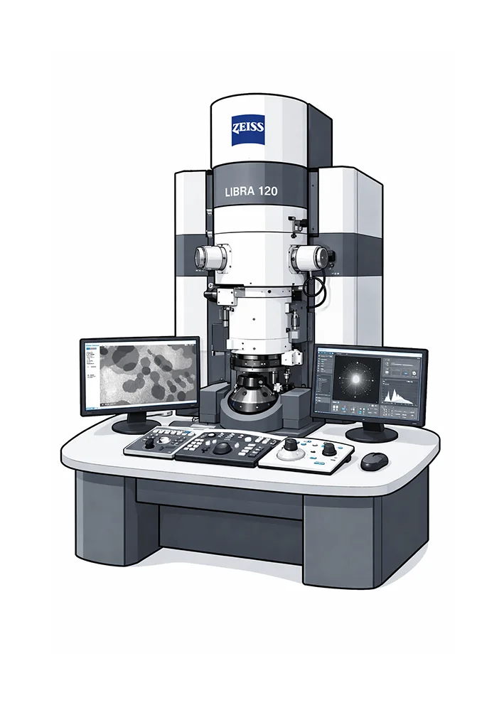
- High-resolution TEM imaging with a spatial resolution up to 0.34 nm and an information limit of 0.20 nm
- Wide magnification range for both low-magnification overviews and high-resolution structural studies
- Analytical capabilities including electron energy loss spectroscopy (EELS) and electron spectroscopic imaging (ESI)
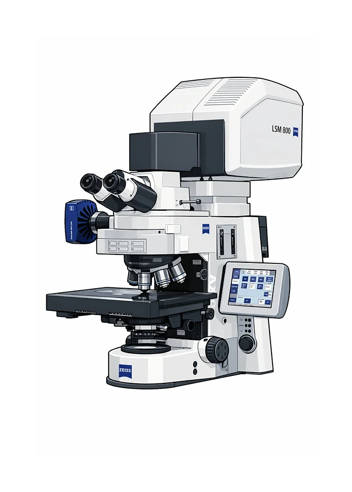
- Multi-color laser scanning confocal imaging powered by four diode lasers for flexible and precise fluorescence excitation
- Airyscan module that maximizes signal collection to enhance resolution, sensitivity, and spatial detail
- Advanced imaging workflows: high quality 3D Z-stacks, colocalization analysis, large-area tiling, and quantitative image analysis using Zeiss ZEN software
- Extensive antibody portfolio, with more than 400 antibodies established and validated for confocal imaging