About us

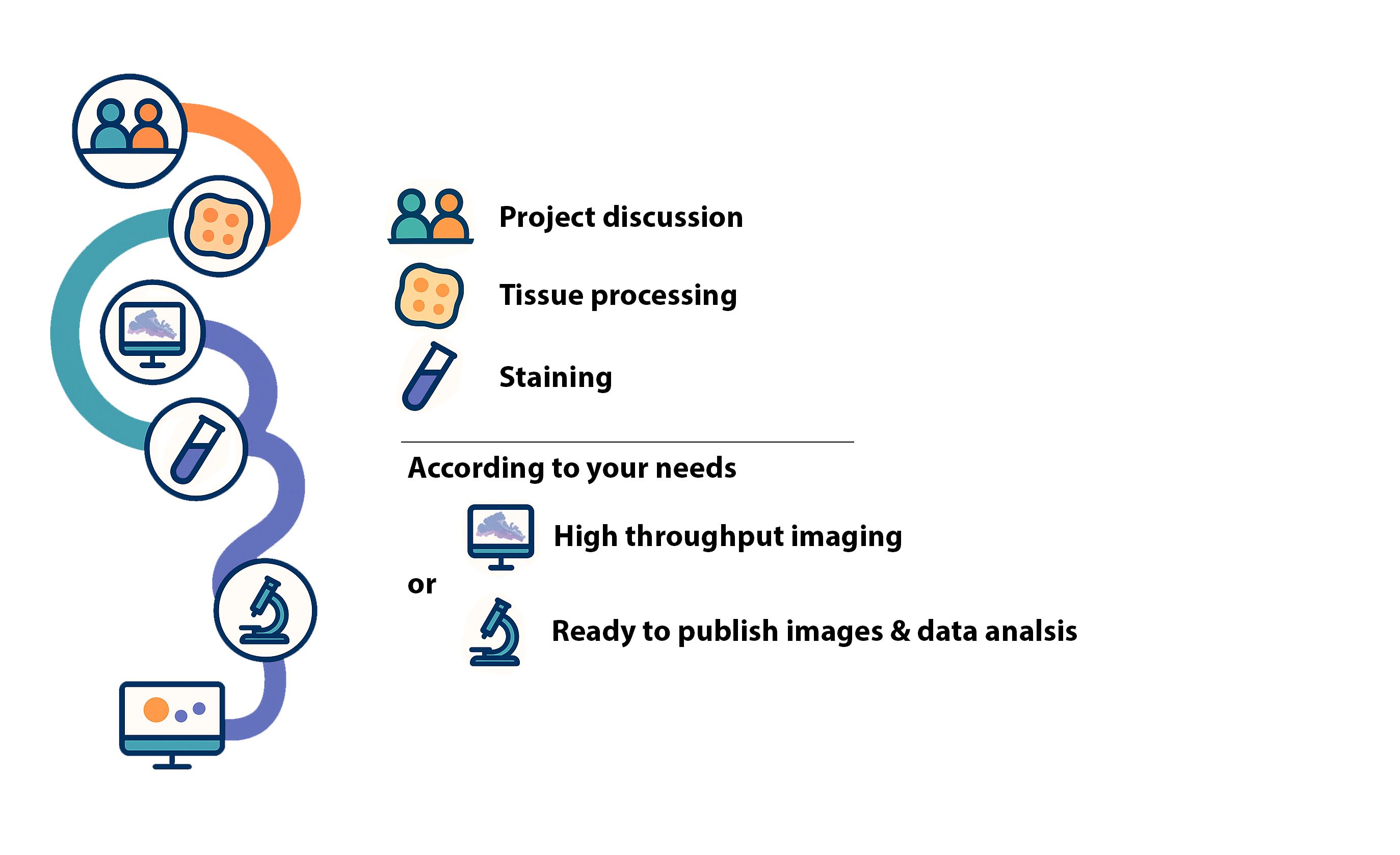
At our Core Facility, two specialized labs – H3 Tissue Imaging and Mouse Pathology – work hand in hand to provide expert support in tissue processing, imaging, and analysis.

Tissue-based analyses are essential across research fields, from fundamental to clinical studies. Histology enables the identification of structures, cell types, and target proteins through immunohistochemistry, aiding in protein localization and expression analysis.

We ensure quality control at every stage—from sample preparation to data analysis—for reliable, reproducible results. Our project consulting supports study design, technology selection, and data interpretation to efficiently address your scientific questions.


Services

Model evaluation
Selection of appropriate biological models


Study design
Experimental design consultation (e.g. control groups, replicates).


Timeline & milestone planning

Budget & resource planning
Cost estimation for histology, imaging, analysis, etc.


Sample preparation
Development of custom SOPs tailored to project-specific needs.

Support for tissue dissection, perfusion, and fixation to ensure high-quality samples for histology.

Tissue dehydration and embedding


Sectioning of FFPE (formalin-fixed paraffin-embedded) or fresh frozen tissue

Custom sectioning options:
Defined thickness
Serial or step sections
Sterile sections (e.g., for microbiology)
Preparation for advanced techniques (e.g., tissue microarrays)

Standard stains (H&E), special stains (PAS,etc.), immunohistochemistry (IHC)

Over 150 (human and murine) established protocols with reproducible, reliable results. Individual antibody establishment.

High-throughput, high-quality histology with state-of-the-art automated systems.


Whole slide imaging under brightfield conditions with 20x and 40x magnification options and Z-stack imaging for high-resolution images suitable for analysis.


High-resolution images suitable for analysis

Long-standing experience in the histopathological interpretation of mouse models.


 Detailed findings report

Publication-ready images


Support with manuscript preparation and revisions


Teams

H3 Tissue Imaging

Specialized in high-throughput
tissue processing and staining

Adresse: Otfried-Müller-Straße 37
72076 Tübingen

Telefonnummer: +49 7071 29-61456

E-Mail-Adresse: Jenny.Hetzer@med.uni-tuebingen.de

Einrichtung: How to find us


Prof. Dr. Mathias Heikenwälder

Head of Core Facility Histology

Jenny Hetzer

Technical Head

Dr. Suchira Gallage

Data Scientist

Sabine Schäfer

Technician

Mouse Pathology

Comprehensive histology services
from sample to publication

Adresse: Liebermeisterstraße 8
72076 Tübingen

Telefonnummer: +49 7071 29-84412

E-Mail-Adresse: Irene.Gonzalez-Menendez@med.uni-tuebingen.de

Einrichtung: How to find us


Prof. Dr. Leticia Quintanilla de Fend

Deputy Head of Core Facility Histology

Dr. Irene Gonzalez-Menendez

Scientist

Helga Pertsch

Technician

Equipment

Sample Processing

Histological sample preparation is essential for studying tissue morphology and structure. The process involves fixation, tissue processing, embedding and sectioning. Two common approaches are cryosectioning (Kryo) and formalin-fixed, paraffin-embedded (FFPE) processing.

Images are courtesy of Leica Biosystems GmbH and SLEE medical GmbH.

Leica Histocore Pegasus Plus

Used for efficient tissue processing, including fixation, dehydration, and paraffin infiltration.

Leica Histocore Arcadia Embedding Station

Used for embedding tissue samples in paraffin before sectioning.
Consists of a heated embedding module and a cooling plate for efficient workflow.

Leica Histocore Peloris 3

Used for efficient tissue processing, including fixation, dehydration, and paraffin infiltration.

SLEE Medical MPS/P

Used for embedding tissue samples in paraffin before sectioning.
Consists of a heated embedding module and a cooling plate for efficient workflow.


Sectioning FFPE

Sectioning of FFPE tissue blocks with customizable thickness, suitable for histology, immunohistochemistry, or molecular analysis. Step cuts for assessing full tissue depth or serial sections for consistent cell layer targeting available.

Images are courtesy of Leica Biosystems GmbH, Epredia Inc. and Thermo Fisher Scientific Inc.

Leica Microtome Histocore Autocut

Rotary microtome with Automated, Semi-Automated, and Manual Modes.

Epredia HM325 STS

Microtome with section transfer system for high-throughput.

Thermo Scientific Microm HM3555

Fully automated, high-precision rotary microtome particularly suited for routine paraffin sectioning.


Sectioning Kryo

Cryosectioning of frozen tissue with adjustable thickness, ideal for histology, immunofluorescence, or molecular applications. Unlike FFPE sectioning, cryosectioning preserves native protein structure and enzyme activity, making it better suited for antigen-sensitive assays.

Images are courtesy of Leica Biosystems GmbH.

Leica Cryostat CM3050S

Adjustable deep temperature cryostat used for fresh frozen tissue.

Standard Staining

Standard staining is a fundamental step in histological analysis used to visualize cellular and tissue structures. It typically involves the application of routine dyes, such as hematoxylin and eosin (H&E), using manual or automated protocols to ensure consistent and interpretable results.

Images are courtesy of Leica Biosystems GmbH and Sakura Finetek GmbH .

Leica Histocore Spectra Workstation & Coverslipper

Combines automated staining and coverslipping in one workflow

High-throughput capacity
Processes large slide volumes efficiently

Consistent quality
Ensures standardized, reproducible results

SAKURA Tissue-Tek Prisma Plus

Automated slide staining
For routine H&E and special stains

Customizable protocols
Supports standardized and flexible workflows

High-throughput processing
Fast and efficient staining

Immunohistochemistry Staining (IHC)

Immunohistochemistry is a widely used method for detecting specific proteins in tissue sections using antigen–antibody interactions. The technique enables spatial analysis of molecular markers and is commonly performed on automated platforms to ensure reproducibility and high-throughput processing in research settings.

Images are courtesy of Leica Biosystems GmbH and Roche Diagnostics.

Leica BondMax

Automated staining system for IHC and ISH

Consistent results
Delivers high-quality, reproducible staining

Leica Bond RX

Automated research staining
Supports IHC, ISH, and multiplex assays

Flexible protocol control
Open access for customized staining workflows

High reproducibility
Ensures consistent results for complex research applications

Ventana Discovery Ultra

Automated staining platform
Supports IHC, ISH, and multiplex applications

Open system design
Allows full protocol customization and reagent flexibility

High-throughput capability

Imaging plays a critical role in histological analysis, enabling detailed examination of tissue architecture, cellular organization, and marker expression through state-of-the-art microscopy.

Images are courtesy of Leica Biosystems GmbH, Hamamatsu Photonics K.K and Roche GmbH.

Leica Aperio AT2

High-throughput scanning
Scans up to 400 slides with minimal user intervention

High-resolution imaging
Produces detailed whole-slide images for precise analysis

Digital workflow integration
Enables efficient storage, sharing, and image analysis

Hamamatsu Nanozoomer RS

High-speed slide scanning
Rapid digitization of brightfield slides

High-resolution imaging
Delivers sharp, detailed whole-slide images

Roche Ventana DP200

Fast slide scanning
High-speed digitization for brightfield histology

High image quality
Produces sharp, diagnostic-grade whole-slide images

FAQ

Please contact us to meet together and get an introduction to your research project. We will give you recommendations regarding the experimental design, number of animals and tissues needed for analysis. We will also provide advice on sample preparation.

We will provide you with an official quote for the analysis of your samples.

You can either send them to us or bring them personally. In any case, please contact us beforehand to coordinate date and time, and give you some guidelines for proper transportation.

We need to know the details of your project to estimate the time required for it. In our first meeting, we will be able to give you a feasible time period based on your project and our workload. You will also be asked whether there is a deadline for publication, grant proposal or doctoral thesis.

We have a range of established antibodies routinely used for immunohistochemistry (IHC) and immunofluorescence (IF) on various tissue types. We are happy to provide information about available and validated antibodies upon request. Please contact us with your area of interest, and we will let you know which antibodies are currently established in our lab.

H3: You will get the raw imaging data.
MP: You will receive a detailed report of our findings as well as ready to publish images. We will also support you through manuscript preparation and revision.