
Degenerative diseases and acute conditions, such as tumors of the locomotor system, demand effective regeneration of bone and soft tissue defects. To achieve healing, both transplants and advanced tissue-engineered constructs are utilized. Our cell biology laboratory delves into the molecular underpinnings and mechanisms driving tissue regeneration. This field presents substantial opportunities for orthopedics, including therapies for repairing joint cartilage and bone defects with cultivated tissue, and addressing joint inflammation through targeted active substances.
Laboratory of Cell Biology
Contact us
Orthopaedic Cell Biology Research Laboratory Derendingen
frontend.sr-only_#{element.icon}:
Waldhörnlestraße 22
72072 Tübingen
frontend.sr-only_#{element.icon}: +49 7071 29-80479
|
Lab Manager |
Management
Group Leader
Dr. phil (PhD) Marina Danalache
Group Leader Cell biological research laboratory
Publications: Publications
Research assistant

Postdoc
Publications: Publications
Our laboratory introduces itself


When you play the video, data is transmitted to YouTube. Further information can be found in our privacy policy.
Current research projects
Innovative In Vitro Approaches to Cartilage Regeneration
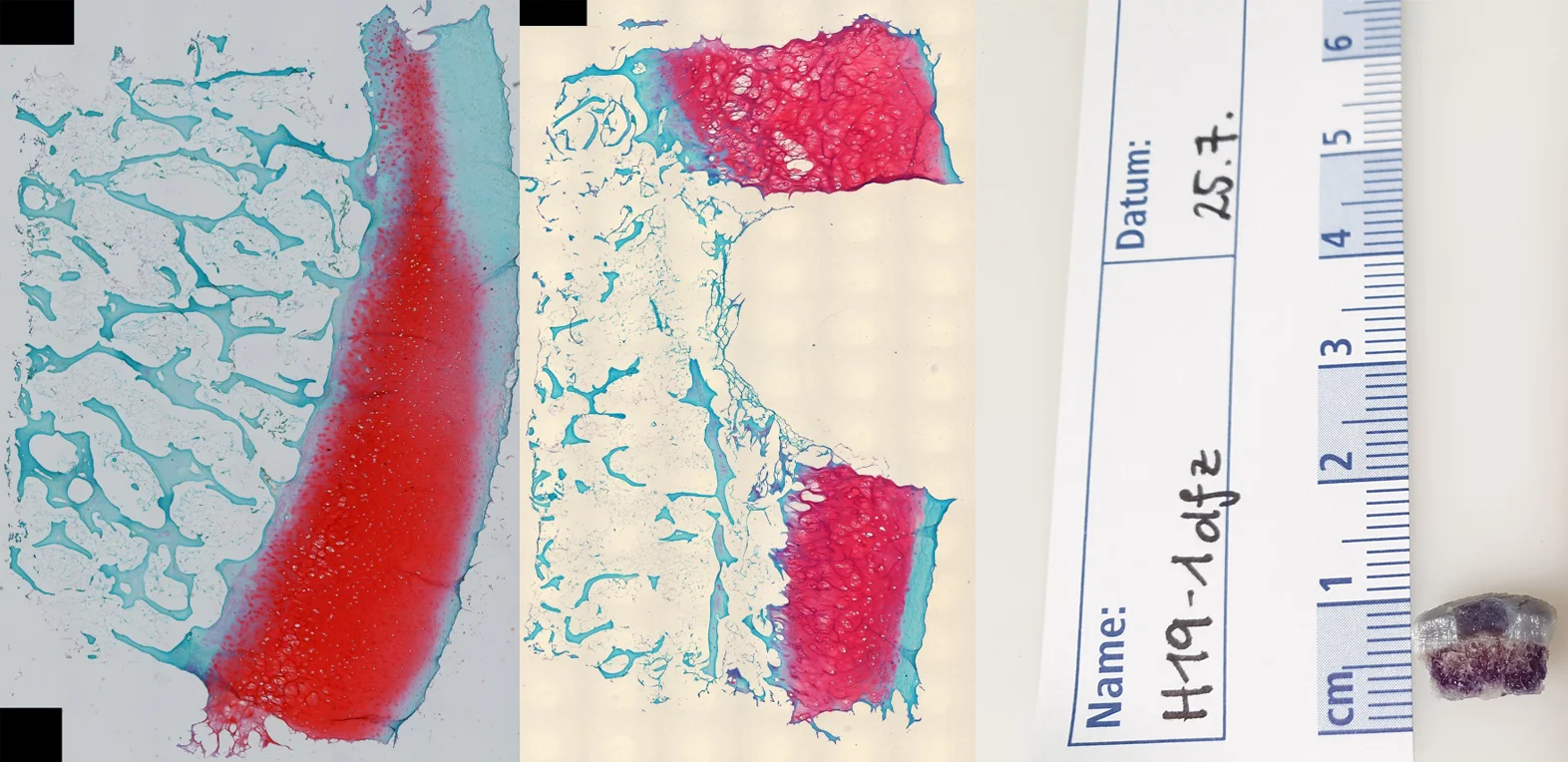
Joint articular cartilage, due to the lack of a perichondrium, is considered to have limited regenerative capacity and, as a result, can be worn down by injuries or misalignment leading to osteoarthritis. In a realistic osteochondral model, artificial regeneration matrices are being studied for their ability to support new cell ingrowth and functionality. The aim is to find long-term alternatives to artificial joint replacement.
What are the impacts and effects of combining chemotherapeutic agents with radiation therapy on tumor signaling pathways?
In cell differentiation, as observed in tumors, multiple signaling pathways are involved. We analyze how the overexpression of specific genes, which influences cellular protein synthesis, correlates with observed changes in cellular biomechanical properties and the effects of radiation therapies. This comparison allows us to draw conclusions about the metastatic potential (cell migration) of these cells.
Chondrocytes and the Pericellular Matrix: Dynamics and Interactions
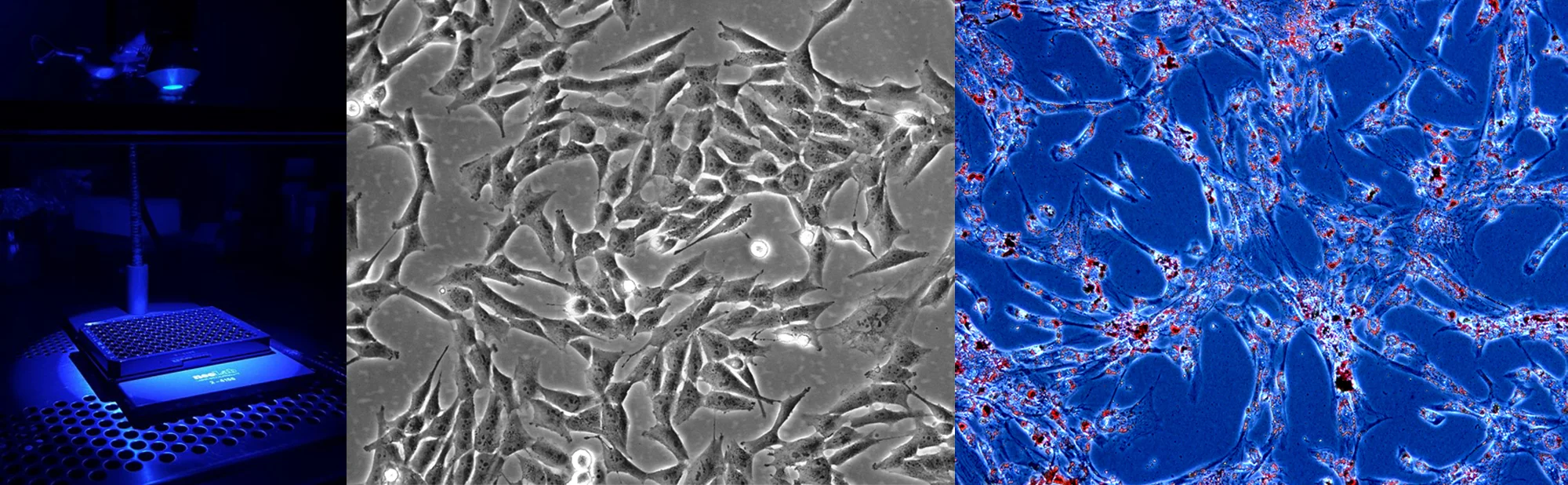
Joint cartilage endures significant compressive and shear forces during loading. Within this cartilage, chondrocytes are scattered throughout an extensive extracellular matrix, which is engineered to handle these high demands with minimal friction. Surrounding each chondrocyte is a specialized form of extracellular matrix known as the pericellular matrix. This cocoon-like layer not only offers mechanical protection but also appears to facilitate coordinated signaling from the extracellular matrix into the cell. We delve into the intricate composition and functional roles of this pericellular matrix to better understand its impact on cartilage health and functionality.

Microbiomechanics: Unveiling the Dynamics of Tissue Matrix
Imaging techniques typically capture qualitative morphological changes in tissues, which are then correlated with quantitative data from biochemical analyses. In our lab, we take this a step further by measuring mechanical changes in tissues and cells at the microscopic scale. This approach enables us to directly translate structural observations into functional insights.
Decoding Biochemical Signaling and Mechanotransduction in Osteoarthritic Cartilage
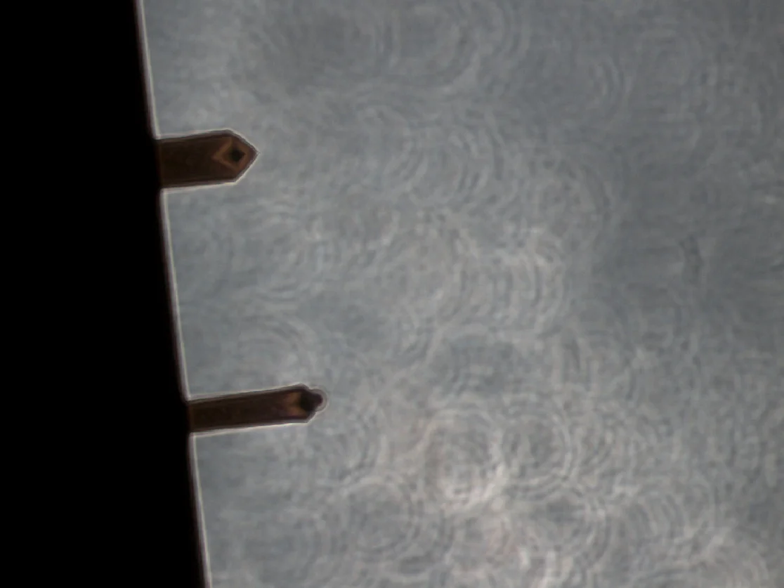
The pericellular matrix (PCM) is crucial for mechanotransduction in chondrocytes. To explore how a compromised PCM affects chondrocyte biochemical signaling, we examine calcium signaling dynamics. By enzymatically isolating chondrocytes and partially degrading their PCM with matrix metalloproteinases, we can probe the impact on cellular responses. Mechanical stimulation is applied using atomic force microscopy (AFM), and calcium influx is assessed through fluorescence microscopy. Our aim is to elucidate the PCM's specific role in mediating cellular mechanotransduction.


The Impact of Specific Calcium Channels on Osteoarthritis Pathogenesis
As a load-bearing tissue, articular cartilage houses a range of mechanosensitive calcium channels that react to mechanical forces and loading. We are investigating the roles of ion channels like Piezo-1, Piezo-2, and TRPV-4 in calcium signaling within chondrocytes. By comparing the expression of these channels in healthy versus osteoarthritic cartilage, our goal is to identify the key signaling pathways that drive the onset and progression of osteoarthritis.

Cortisone: Friend or Foe?
We investigate the dual role of glucocorticoids (Cortisone) in knee joint tissue regeneration and degeneration.

Theses
Doctoral theses and scientific dissertations in the cell biology research laboratory
We are looking forward to welcoming motivated colleagues who enjoy exploring our exciting research area with us in our international team.
In principle, there is the possibility to do a scientific (Dr. sc. hum., PhD) or medical doctorate (Dr. med./Dr. med. dent.) or to write a scientific thesis (Bachelor/Master thesis) with us.
The scientific doctorate is embedded in the graduate college of the medical faculty and is scheduled to last three years. Bachelor's and Master's theses are carried out according to the regulations of the sending university. In the case of medical doctorates, a semester off is necessary for the success of the thesis in order to ensure the high scientific level of an experimental medical doctorate. If planned in time, the application for an IZKF scholarship can also be discussed here.
Teaching commitments
Teaching commitments
We are involved in the following courses of the medical Faculty of Tübingen:
- compulsory election (medical students)
- block internship (Medical technology)
Contact for PhD/Dr.med/MSc/BSc- Projects:
Certificates and Associations

Focus: Top National Hospital 2026


Stern: Germany's Outstanding Employers in Nursing 24/25


Quality partnership with the PKV

Family as a success factor

Pension provision for the public sector
