
Bettina Weigelin is researching microscopic methods in Germany's only oncology cluster of excellence, which can be used to observe living tissue at cellular level, among other things. This contributes to understanding immunotherapies in particular - and to improving treatment.
Medicine is often still a black box. This is particularly true for research, but also for everyday clinical practice. You administer an active substance - and wait for the effect it brings about. If the hoped-for effect does not occur, the administration is varied, for example with a different concentration or a different active ingredient. Trial and error.
How much better would it be if you could observe what happens in the time between administration and the effect? If, for example, you could watch the T cells at work - how they attack tumor cells, for example, and what difficulties they have to contend with?
Making the invisible visible

It is precisely this look behind the scenes that Prof. Dr. Bettina Weigelin is trying to provide. The Professor of Preclinical Imaging of the Immune System and group leader at the Werner Siemens Imaging Center (WSIC) at Tübingen University Hospital is conducting research using two innovative imaging techniques with cellular resolution: Light sheet microscopy and intravital microscopy. "Thanks to their insights at the cellular level, the new methods can be an ideal complement to MRI and PET and provide us with valuable information about the specific mechanisms of action of treatments, among other things," says Weigelin. Her work is also part of Germany's only oncology cluster of excellence, the "Image-Guided and Functionally Instructed Tumor Therapies (iFIT)". The aim here is to better understand why certain oncological therapies work for some patients and not for others. And how could the therapy be changed to make it more effective?

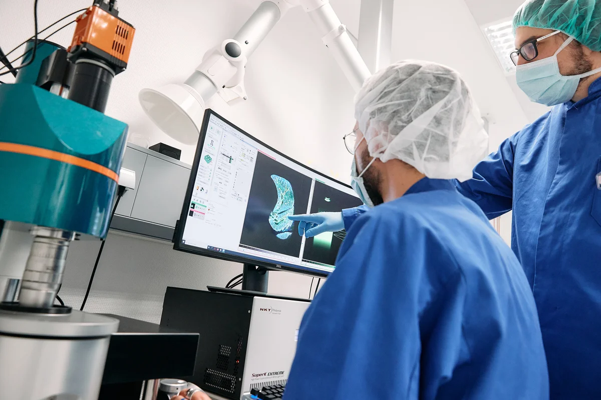
Bettina Weigelin shows what this looks like on the screen. Intravital microscopic images of a malignant melanoma in a mouse appear here. "We can use fluorescent proteins to make certain cells in tissue samples visible," says the professor. "Here we see a tumor around which T cells have arranged themselves in a ring structure. They specifically attack the tumor cell as part of an immunotherapy." Next image: The T cells have disappeared, but the tumor is still there. Bettina Weigelin explains why: "We were able to show that tumor cells continue to exist when they are not attacked by a large superiority of T cells. Instead, the wounded tumor cells apparently change, they become resistant, temporarily stop dividing - and then continue to grow after a certain period of time." Fatal for the patients.
Bettina Weigelin and her team therefore researched how this problem could be solved and realized that it occurs much less frequently with a larger number of T cells. "We suspect that some immunotherapies are not effective if the number of T cells is too small," says Weigelin. She goes on to specify what it takes to sustainably combat tumor cells as part of immunotherapy: "A larger number of T cells alone is only one factor; the density of the T cells is also important: They should not be evenly distributed in the tumor, but should be in higher density in a specific region." Important findings that can be used to further improve therapy.
The microscopic methods used by Weigelin and her team are not only useful for the evaluation and better understanding of cell-based immunotherapies; the effect of other immunotherapies, such as checkpoint inhibitors, can also be examined and better understood with the help of microscopy.
The fact that living tissue can be observed at all at cellular level is an innovation of recent years that is still being further developed - this is all the more appealing for researchers like Bettina Weigelin. The intravital microscope in Tübingen cost around one million euros and takes up several square meters. The room in which it is located can only be entered with disposable overalls.
The light sheet microscope, on the other hand, cannot be used to examine living organisms, but it can be used to examine intact mouse organs in layers. The aim is to better understand metastasis and the treatment of metastases. "If we stain certain cells in the tissue, we can make the remaining tumor cells visible after treatment. We understand where how many tumor cells are left and can compare this result with combination therapies, for example," explains Bettina Weigelin.
The observations at the cellular level can also be derived from macroscopic imaging procedures that are widely used in the clinic, such as PET and MRI.
To stay with the tumor surrounded by T cells: "Thanks to our microscopic procedures, we have a better understanding of the basic processes of how and where the T cells arrange themselves around the tumor - which in turn makes it easier for us to interpret and understand certain patterns in the MRI," says Weigelin. The findings from the cellular level therefore help to improve the interpretation of MRI images in many cases. A further step from "knowing that it works" to "understanding exactly how it works". Bettina Weigelin and her team are slowly finding more peepholes to look into the black box.