330

Address: Calwerstr. 7
72076 Tübingen


E-mail address: face@​med.​uni-​tuebingen.​de


frontend.sr-only_#{element.icon}: +49 7071 29-83799


Craniosynostoses - growth disorders of the skull

Craniosynostoses

Growth disorders of the skull

A craniosynostosis is a premature ossification of one or more of the so-called cranial sutures, which is accompanied by a circumscribed or general growth disorder of the cranial skeleton. The growth impulse comes from the rapidly enlarging brain, not from the bone itself.

There are four main cranial sutures (frontal, coronal, sagittal and lambdoid sutures) which, with the exception of the frontal suture, which ossifies towards the end of the first year of life, do not normally close until the second to third decade of life. The open cranial sutures during childhood ensure that the cranial bones are displaced during birth and also enable rapid cranial growth due to the very fast growing brain in the first year of life.

If one or more cranial sutures are closed prematurely, the growth of the skull is inhibited, resulting in a characteristic deformation of the skull.

Father lies with child on chest
The general goal of therapeutic measures for craniosynostosis is to eliminate, mitigate, or prevent as many of the negative consequences of synostosis as possible, thereby creating optimal conditions for the child's development.

Interdisciplinary consultation

Contact

Interdisciplinary consultation
for Craniofacial Anomalies

frontend.sr-only_#{element.contextual_1.children.icon}: Neuropediatric Outpatient Clinic,
Children's Hospital C3
Hoppe-Seyler-Str. 1
72076 Tübingen, Germany


Directions

Thursday 3-5 pm
1x monthly

+49 7071-29-83806


Departments involved:
Neuropediatrics
(OA Dr. Jan Kern, Dr. Karin Haas-Lude)
Neurosurgery
(Prof. Dr. Martin Schuhmann)
Oral and maxillofacial surgery
(Prof. Dr. Dr. Michael Krimmel)


More about the Neuropediatric Outpatient Clinic

Referring clinics and practices

Contacts and contact persons

Learn more

Therapy

The general goal of therapeutic measures for craniosynostosis is to eliminate, mitigate or prevent as many of the negative consequences of synostosis as possible, thereby creating optimal conditions for the child's development. Depending on the form and characteristics of the synostosis, aesthetic aspects or reasons such as lowering of increased pressure in the head or protection of the eyes by enlarging the eye socket are in the foreground.

Depending on the form of synostosis, there are two surgical principles, which are performed in joint work by pediatric neurosurgery and oral and maxillofacial surgery.

Passive remodeling

In passive remodeling, as used for sagittal suture synostosis, for example, the correspondingly ossified cranial suture and the adjacent bones of the cranial calvaria are removed. Within a few months, the bone is remodeled by the body (called reossification) and the skull is remodeled in the interval in between according to the shaping force of the growing brain. This operation should be performed between the 5th-6th month of life, because at this time of maximum brain growth the internal pressure in the head is highest and the reossification also occurs without problems. Surgery at an earlier time, e.g. at 3 months of age, requires additional treatment with a helmet to support the reshaping. This early endoscopically assisted therapy offers advantages especially in metopica and coronary suture synostoses, as a later, reshaping and major surgery can often be avoided.

Active remodeling

Active remodeling is used, for example, in frontal suture/metopicasynostosis or isolated coronal suture synostosis at the age of 10-12 months. In this case, the skull bone in the area of the forehead and the roof of the orbit, including the ossified suture, is removed, divided into suitable segments, actively remodeled and then reassembled in the desired shape and fixed to the base of the skull. This improves the deformity in the area of the forehead and ocular skeleton and, in the case of syndromal craniosynostoses, also creates space in the head.

In some synostoses, a backward extension of the skull is also applied.

The most important risk of these operations is relevant blood loss, which is mainly due to the large wound area. In this case, the administration of blood reserves and clotting factors is often necessary despite all blood-saving surgical techniques and measures taken during the operation as a rule. After the operation, the children spend one night in our pediatric intensive care unit to ensure optimal care and monitoring of blood values. The total inpatient stay is between 4-7 days, depending on the type of surgery and the age of the child.

The probability of a craniosynostosis recurring after a successful operation, i.e. the space for the brain becoming too small so that a second operation is necessary, is a maximum of 10% for isolated synostoses and at least 60% for syndromes with many affected sutures.


Follow-up

After surgery, children are followed up regularly for years in our craniofacial consultation by a team of pediatric neurosurgeons, oral and maxillofacial surgeons, and neuropediatricians to detect any recurrences early.

Forms, frequency and causes of craniosynostosis

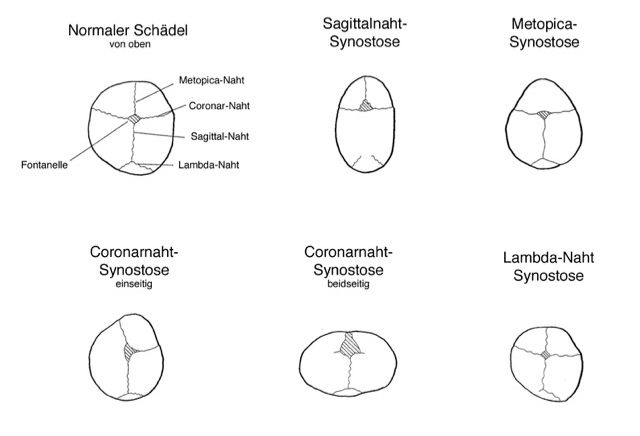
Scientific illustration
Premature suture closure with resulting characteristic head shape compared to the normal skull shape with open cranial sutures. The closed suture is no longer visible in the respective image.

Sagittal suture synostosis results in an elongated, narrow skull shape with a protruding occiput and prominent forehead (so-called scaphocephalus) due to the altered direction of growth. The fontanel is often narrow or completely closed and a bony ridge can be felt in the area of the sagittal suture.

Metopicasynostosis leads to a tapered head shape in the forehead area, often with a palpable bony ridge at the metopic suture. The shape of the forehead may not develop normally and there are often retractions in the area of the temples or over the lateral region of the eye sockets. As a compensatory reaction, increased growth of the skull in the posterior region can often be observed. All changes together lead to a classic triangular head shape (so-called trigonocephalus), especially when viewed from above.

In unilateral coronal synostosis, an asymmetry of the forehead and face develops (so-called facial scoliosis) with flattening of the forehead and eye socket on the affected side and bulging of the forehead on the opposite side (so-called anterior plagiocephalus). The eye on the affected side often appears larger and the nose may deviate towards the opposite side. Bilateral coronal synostosis is very rare and leads to increased skull growth in width (brachycephalus). A very high and flat forehead develops and the eye sockets may be reduced in size. The skull often grows upwards in a compensatory manner (so-called turricephalus).

In lambdoid synostosis, there is a unilateral asymmetry of the back of the head (so-called posterior plagiocephalus) with flattening on the affected side and compensatory protrusion on the opposite side. Viewed from the front, asymmetry of the ears may be noticeable. The main differential diagnosis to the very rare lambdoid suture synostosis is the so-called and relatively common "positional plagiocephalus", in which the lambdoid sutures are open.

Frequency

The incidence of primary craniosynostosis is 1/2500, with isolated sagittal suture synostosis accounting for about 50% of all cases. The most common syndromes associated with craniosynostosis are Apert, Crouzon, Muenke and Saethre-Chotzen syndromes. They each occur only once in about 30,000-80,000 births.

Causes

The cause of isolated synostoses is still unknown. A familial accumulation of isolated sagittal and frontal synostoses of around 5% suggests a hereditary cause in some families, but the genetic defect (location of the genetic alteration) is still unknown. For the most common syndromes, however, the genetic alterations are known and the diseases are inherited autosomal-dominantly, i.e. with a probability of 50%.

Symptoms and clinic

The consequences of synostoses, skull deformities and other possible malformations that can occur in connection with a syndrome are

  • Aesthetically problematic cranial deformities, which can lead to social exclusion and stigmatization
  • Development of increased pressure in the head - "intracranial pressure"
  • An eye socket that is too shallow
  • Underdevelopment of the midface
  • Accompanying malformations of the brain, extremities and internal organs
  • Developmental delays (inconsistent, depending on the clinical picture)

Very conspicuous skull deformities often have a disfiguring effect and can lead to social exclusion in later life. Even with isolated suture synostosis, further skull growth is likely to lead to an increase in skull deformities, which can stigmatize and thus impair the affected child. For this reason, the aesthetic aspect is particularly important in isolated suture synostosis.

If the brain develops normally, the growth inhibition of the skull caused by suture synostosis can lead to an increase in intracranial pressure. The extent of this increase in intracranial pressure depends primarily on the number of sutures affected and the time of ossification.
Recent studies at our clinic have shown that premature ossification of a single suture, particularly the sagittal suture, can also lead to more pronounced increases in intracranial pressure in up to 80% of children. It is unclear whether and, if so, to what extent this impairs brain development. The occurrence of a more severe increase in intracranial pressure ("intracranial pressure") with the development of classic "intracranial pressure symptoms" is very rare in the case of isolated suture synostosis, but more common in the case of closure of several sutures. There is evidence in the literature that children who were operated on earlier for sagittal suture synostosis (before the age of 6 months) had on average better cognitive function after 10 years than children who were operated on later (after 12 months).

Severe forms of craniosynostosis, as seen in syndromes, may be associated with underdevelopment of the orbits and midface. An underdeveloped and shallow eye socket can lead to incomplete closure of the eyelids with strongly protruding eyes and thus endanger the cornea through dehydration and inflammation. Underdevelopment of the midface can lead to constriction of the airways, which can result in sleep apnea syndrome at night. Other problems that can be associated with an underdeveloped midface include Middle ear infections and hearing loss, incorrect jaw relation with the upper jaw being set back, lack of space for the teeth and impaired chewing function.

In syndromes, further malformations must be expected, such as developmental disorders of the brain, inner ear hearing loss, fusion of fingers and toes (so-called syndactyly) and cleft palate, slipping of the cerebellum due to a too small head into the spinal canal (Chiari 1 formation) or an obstruction of blood outflow from the skull.

Diagnostics and differential diagnosis

Since craniosynostoses are associated with a typical skull shape, the assignment to a specific synostosis is usually a so-called visual diagnosis for experienced physicians. In most cases, no further diagnostic imaging is necessary. Ultrasound examination (sonography) of the cranial sutures can reveal the closure or openness of the 4 large cranial sutures without radiation exposure. If there are still unanswered questions, the next step is a simple X-ray of the skull or a modern, extremely radiation-reduced computer tomography of the skull bones only.

The most important other "disease" (differential diagnosis) in the unilateral flattening of the back of the head is the so-called positional deformity of the skull (positional plagiocephalus), which is the most common skull deformity of all. It occurs in the first few weeks of life due to the child's preferred position on one side of the head and usually improves with positioning measures using special pillows, physiotherapy/osteopathy and then spontaneously with increasing prone positioning of the child and upright posture without the need for therapy. It is important that positioning measures and physiotherapy/osteopathy begin early in the first few months of life. Affected children often have a normal head shape at birth, whereas children with craniosynostosis may already be conspicuous at birth due to their unusual head shape. More extreme head deformities caused by positioning can also be treated with so-called helmet therapy, which should be initiated at the latest in the 5th month of life in order to be effective.

Children with suspected or confirmed craniosynostosis are seen in the interdisciplinary craniofacial consultation in the children's clinic (link) by doctors from neuropediatrics, pediatric neurosurgery and oral and maxillofacial surgery. In addition to visual diagnostics, the cranial sutures can be visualized with the help of sonography at this appointment. To assess any accompanying "intracranial pressure", sonography of the optic nerve sheath diameter (ONSD) can also be performed.

Certificates and Associations

Go to the main content