
Research on fetal programming revealed that intrauterine influences and prenatal experiences have an effect on the development of a child that even persists until adulthood. In order to investigate cognitive development, so far, indirect methods as observation of fetal behavior in response to auditory, visual and vibroacoustic stimulation have been used due to the inaccessibility of the fetus. Well protected by the maternal abdomen and secluded from the external environment, there was no way to directly investigate the developmental processes of the fetal brain without touching the integrity of the uterine environment.
fMEG
Head of research group

Prof. Dr. rer. nat. Hubert Preißl
Institut für Diabetesforschung und metabolische Erkrankungen des Helmholtz Zentrum München an der Universität Tübingen
Publications: Publications
Fetal Magnetoencephalography
Fetal Magnetoencephalography
Recent technical progress makes it possible to respect the integrity of the body as well as directly access brain activity. The MEG technology (Magnetoencephalography), approved in decades of adult research, with its advantages of completely non-invasive recordings of brain signals and superior temporal resolution, has been combined in a new device that takes into account the demands arising from fetal and neonatal measurement requirements.

The new device, referred to as Fetal Magnetoencephalography (fMEG, Preissl et al. 2004), enables to record fetal auditory and visual evoked fields, spontaneous activity, and as a by-product, fetal heart rhythm parameters, which all are detectable from the surface of the maternal abdomen. The latter is interesting in terms of clinical issues, since fluctuations of the heart beating reveal the status of the autonomous nervous system (ANS). Thus, the analysis of fetal heart rate variability helps to conclude fetal development and well-being, to better understand the intra-uterine ANS and, to detect eventual cardiac malfunctions.

The magnetic fields are recorded by 156 highly sensitive sensors (SQUIDs, superconducting quantum interference devices), shaped to perfectly fit the anterior abdominal surface of the mother, from the perineum to the top of the uterus, without exposing mother or child to any radiation. Furthermore, the device is equipped with a custom-made cradle attachment which also allows for keeping track of the child´s development postnatally.
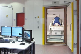
The high sensitivity of the sensors would also pick up natural environmental magnetic fields that are much bigger in amplitude than brain signals, therefore, the device is located inside a shielded room. Near-field biological signals that also cause magnetic interferences like the fetal and maternal heart, uterine contractions, intestinal movements, and motion artefacts have to be extracted in the process of data analysis in order the observe brain responses.
Zwei Systeme weltweit – Tübingen und Little Rock
Two systems worldwide - Tübingen and Little Rock
The first fMEG device, a 151 channel MEG system called SARA (SQUID Array for Reproductive Assessment), was installed in Little Rock, Arkansas, in 2000. Due to a collaboration between the Department of Obstetrics and Gynecology, University of Arkansas for Medical Sciences, and the MEG Center, Tübingen, it was possible to gain insights into auditory and visual processing and the underlying neurophysiological correlates in fetuses and neonates. By recording auditory evoked magnetic fields in response to a single tone, Holst et al. (2005) examined the maturation of the auditory cortex and showed an age related decrease in response latency in fetuses and newborns.
Draganova et al. (2005, 2007) demonstrated fetal brain responses to a tone frequency change using the same SARA system. They performed serial biweekly recordings and showed that it is possible to track the development of fetal brain responses to sound frequency changes starting as early as 28 weeks of gestational age until delivery. The question when and how children start to detect changes in sounds is important, since the ability is a prerequisite to normal cognitive development and related to language learning that already has its roots in the prenatal phase.
Another method towards an early detection of developmental impairments utilises habituation in order to assess neurological integrity and the functioning of the central nervous system. Sheridan et al. (2008) investigated the application of a short-term habituation paradigm in fetuses and newborns. They delivered trains of four light flashes that caused response decrements in amplitude and a significantly increased response latency from the first to the last stimulus. Fetuses showed a response decrement after the first stimulus, although the prenatal visual evoked response rate was low due to a common reason of reduced signal-to-noise ratio for fetal recordings.
In addition to the already existing whole-head MEG system used mainly in adult research, a successor of the SARA system has been installed at the MEG Center in 2008, which is the first fMEG device in Europe and the second worldwide (funded by the DFG and Land Baden-Württemberg).
Certificates and Associations

Focus: Top National Hospital 2026


Stern: Germany's Outstanding Employers in Nursing 24/25


Quality partnership with the PKV

Family as a success factor

Pension provision for the public sector
