Institute of Immunology
Research Group
Immune Monitoring

Contact

Telefonnummer: +49 7071 29-80994

E-Mail-Adresse: cecile.gouttefangeas@uni-tuebingen.de

Adresse: Auf der Morgenstelle 15, 72076 Tübingen

Research

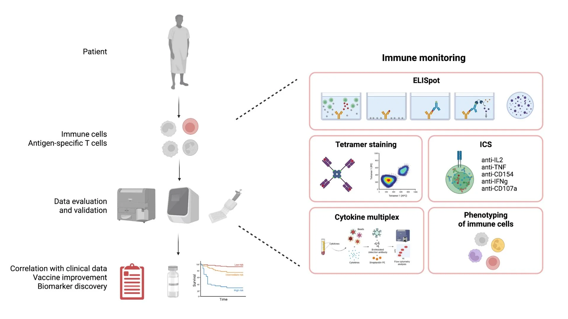
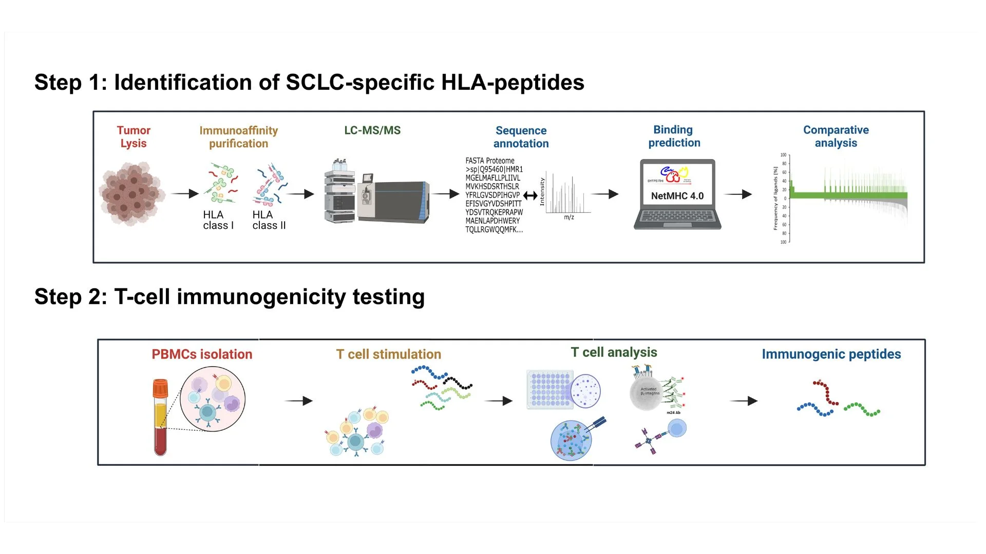
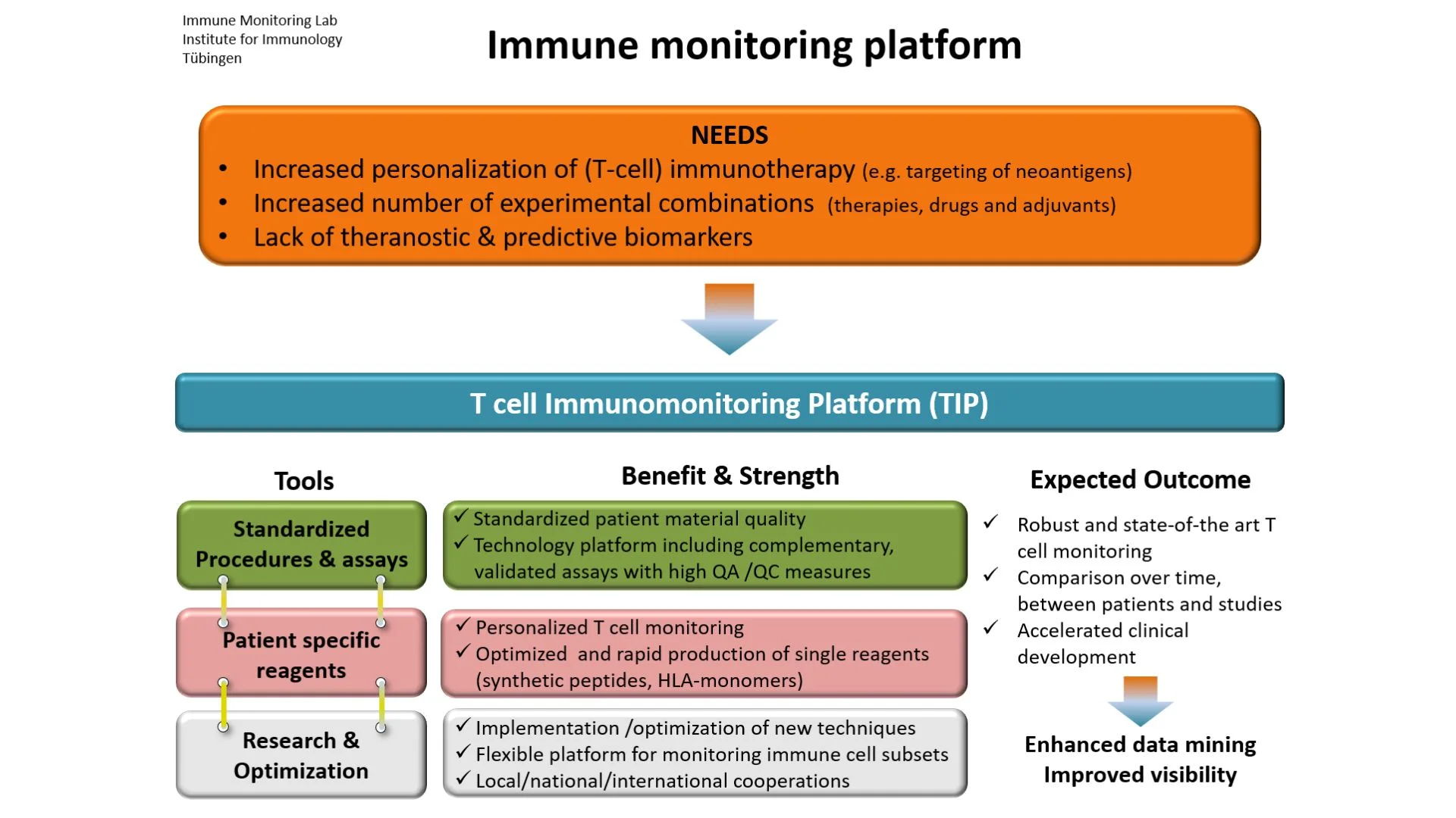
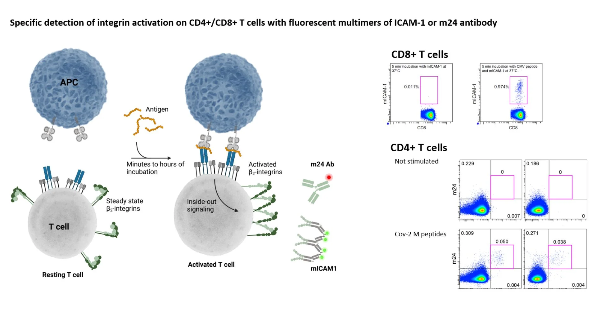
The aim of the immune monitoring team is to contribute to the understanding of immune responses against cancer and to apply this knowledge in immunotherapy. We are focusing on the description of anti tumor T cell responses in cancer patients, either before or during therapy, including immunotherapy. We investigate subsets of T cells at the phenotypic and functional levels, in the blood of cancer patients and also within the tumor tissue. We also assess other immune cell subsets interacting with human tumors and their impact on T cells and clinical course. Finally, we are currently identifying tumor antigens for small cell lung carcinoma.

Key Areas:

  • T cells
  • Cancer
  • Immunotherapy
  • Immunomonitoring

T cell-based immunotherapy of cancer

We are taking part to experimental vaccination studies for cancer patients, for which we are responsible for the full immune monitoring during vaccination. Aim of this in-depth immune monitoring is to assess i) the immunogenicity of the peptides contained in the vaccine, ii) the quality of the vaccine-specific T cells, iii) the impact of further immune cell subsets on the T cell response, iv) the action of the adjuvant, and, importantly, iv) the association between immune cell activation and clinical response (Figures 1&2). 

Figure 1: Workflow for in-depth immune characterization during immunotherapy of cancer. The left panel depicts the major steps between the collection of immune cells (blood or TILs) of patients and the final analysis. The right panel shows our experimental platform with the main in vitro testing methods we are using. Created with Biorender.
Figure 2: Example of the polyfunctional analysis of vaccine-specific T cells in a cohort of prostate carcinoma patients. Bars represent means + 95% CI of specific CD4+ T cells expressing each of the five activation markers or all possible combinations thereof. One example of an intracellular cytokine staining is shown. Schuhmacher et al. Journal Immunother Cancer 2020, 2:e001157

Immunopeptidomics

Our lab is part of the SFB1399 “Mechanisms of Drug Sensitivity and Resistance in Small Cell Lung Cancer” https://www.sfb1399.de

Small cell lung cancer (SCLC) is the most aggressive subtype of lung cancer. It is initially sensitive to chemoradiotherapy and PD-L1 blockade, but in most cases, relapses with no effective treatment option left. SCLC-associated antigens that are recognized by T cells are largely unknown. In this project, we aim at identifying SCLC-associated T cell targets via mass spectrometry-based immunopeptidomics (workflow in Figure 3). Our long-term objective is to define HLA-class I and class-II presented SCLC-antigens that could be used in immunotherapy approaches, e.g. peptide-based vaccination or adoptive transfer.
Figure 3: Flow-chart for identification of T cell antigens in small cell lung carcinoma. The upper panel shows the mass-spectrometry approach for extracting MHC peptides and identifying their source proteins. The lower panel depicts the experimental approach for validating SCLC-specific peptides as T cell targets. It involves both T cell priming and recall experiments with T cells from healthy donors and SCLC patients, respectively. Created with Biorender.

Immune monitoring platform

Immune monitoring platform

Our laboratory is equipped with high quality tools for immune monitoring. We are investigating different immune biomarkers associated with cancer and (response to) therapy (Figure 4: schematic and aims of our immune monitoring platform). We are also interested in developing new “fit for purpose” antibody panels and assays for addressing the phenotype and function of immune cell subsets (Figure 5: a new assay based on the detection of conformational changes of integrins after T cell activation). Our lab was one of the founding members of the CIP/CIMT network and is part of the iFIT Cluster of Excellence.
Figure 4: Immune monitoring is essential from basic to translational research. Our technical platform (TIP) focuses on the characterization of immune biomarkers in cancer patients and during T cell-based immunotherapy.
Figure 5: Assessment of adhesion properties as a T cell monitoring tool. Left figure: following T-cell receptor-mediated stimulation, integrin (LFA-1) activation occurs rapidly through an “inside-out” signaling process which leads to an affinity increase and a clustering of membrane-bound integrins. These can be detected using fluorescent multimers of their ligand ICAM-1 (for detection of antigen-specific CD8+ T cells) or using the monoclonal antibody m24 (for detection of antigen-specific CD4+ and CD8+ T cells). Created with Biorender. Right panel: flow cytometry analysis of T cells after short stimulation with overlapping peptides. Cytometry analysis of the T cell response as detected with m24 Ab staining combined to intracellular cytokine staining (upper dot-plots), or to extracellular activation markers (lower dot-plots). Dimitrov et al. Proc Natl Acad Sci U S A 2018, 115:E5536, Schöllhorn et al. Front Immunol 2021,12:626308, Schöllhorn et al. Front Immunol 2023, 13:1107366.