The PupilPanda project emerged from a series of studies that investigated how perception-based stimuli can be used to study visual function at various physiological levels. The work is based on investigations of the Panda illusion, a visual stimulus that relies on the principle of pulse-width modulation. We were able to demonstrate that the illusion is decoded by the visual system through spatial low-pass filters. This explains why reduced visual resolution can improve the visibility of the embedded panda (Straßer et al., Scientific Reports, 2020). This perceptual property established a direct relationship between the detectability of the illusion and spatial frequency.
Building on this concept, the illusion was initially evaluated as a tool for determining visual acuity. It was found that the detection thresholds systematically vary with spatial frequency and blur. This enables the estimation of functional visual acuity using a perception-based stimulus instead of conventional optotypes (Kelbsch et al., British Journal of Ophthalmology, 2023). These results established the panda illusion as a controllable probe of visual resolution.
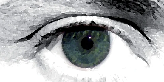
In the next step, the paradigm was extended from psychophysics to objective electrophysiology. In the study “Seeing the Panda,” the illusion was embedded in a visual oddball design to elicit both visual evoked potentials (VEP) and P300 responses (Nikolaidou & Strasser, ISCEV 2025 Conference). This approach enabled the simultaneous investigation of early sensory processing and higher-level cognitive evaluation of the stimulus. The results showed that the amplitudes of VEP and P300 covary with optical blur and spatial frequency. This supports the assumption that refractive and cognitive aspects of visual acuity can be captured within a unified neural framework. Following the demonstration of robust cortical effects, the paradigm was extended to pupillography. In the study “Your pupil knows better: Functional Pupillography with the Panda Illusion” (Nikolaidou, Schedel & Strasser, ARVO 2026 Conference), the illusion reliably elicited event-related pupillary responses. Notably, the pupil dilation patterns differed from the reactions recorded via button press. This suggests that autonomic responses follow stimulus-driven perceptual processing even when conscious perception is inconsistent. These results suggest that pupil dynamics offer an additional, objective window into visual processing.
Previous work:
- Straßer, T., Kurtenbach, A., Langrová, H. et al. The perception threshold of the panda illusion, a particular form of 2D pulse-width-modulated halftone, correlates with visual acuity. Sci Rep 10, 13095 (2020). https://doi.org/10.1038/s41598-020-69952-6
- Kelbsch C, Spieth B, Zrenner E, et al. PandAcuity in pediatrics: a novel clinical measure of visual function based on the panda illusion. British Journal of Ophthalmology 2023;107:582-586.